


![[The AI Show Episode 143]: ChatGPT Revenue Surge, New AGI Timelines, Amazon’s AI Agent, Claude for Education, Model Context Protocol & LLMs Pass the Turing Test](https://www.marketingaiinstitute.com/hubfs/ep%20143%20cover.png)


.png?#)


.webp?#)
.webp?#)


![[Fixed] Gemini app is failing to generate Audio Overviews](https://i0.wp.com/9to5google.com/wp-content/uploads/sites/4/2025/03/Gemini-Audio-Overview-cover.jpg?resize=1200%2C628&quality=82&strip=all&ssl=1)

![What’s new in Android’s April 2025 Google System Updates [U: 4/14]](https://i0.wp.com/9to5google.com/wp-content/uploads/sites/4/2025/01/google-play-services-3.jpg?resize=1200%2C628&quality=82&strip=all&ssl=1)


![Apple Seeds tvOS 18.5 Beta 2 to Developers [Download]](https://www.iclarified.com/images/news/97011/97011/97011-640.jpg)
![Apple Releases macOS Sequoia 15.5 Beta 2 to Developers [Download]](https://www.iclarified.com/images/news/97014/97014/97014-640.jpg)


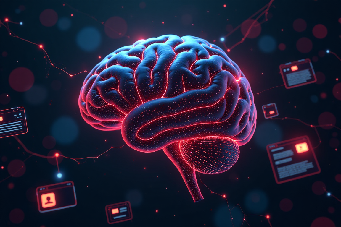
Scientists unveil new wiring diagram tracing millions of connections in a bit of brain tissue
Researchers say they’ve accomplished a feat that was said to be impossible 46 years ago: mapping the cells in a cubic centimeter of brain tissue and tracing their activity. The achievement, documented today in a set of 10 research papers published by the Nature family of journals, is being compared to the Apollo moon shots that were launched more than 50 years ago, and to the drafts of the human genome that were released more than 20 years ago. Scientists from Seattle’s Allen Institute played a key role in the $100 million effort known as the Machine Intelligence from Cortical… Read More


Researchers say they’ve accomplished a feat that was said to be impossible 46 years ago: mapping the cells in a cubic centimeter of brain tissue and tracing their activity.
The achievement, documented today in a set of 10 research papers published by the Nature family of journals, is being compared to the Apollo moon shots that were launched more than 50 years ago, and to the drafts of the human genome that were released more than 20 years ago.
Scientists from Seattle’s Allen Institute played a key role in the $100 million effort known as the Machine Intelligence from Cortical Networks program, or MICrONS. More than 150 researchers worked together through MICrONS to create a detailed 3D map of a cubic centimeter taken from a mouse’s brain — and figure out how the 200,000 brain cells in a speck the size of a coarse grain of sand work together.
“It really has been one of the holy grails of the field from the beginning,” Clay Reid, a senior investigator at the Allen Institute, told GeekWire. “There are many thousands of neuroscientists who study the cerebral cortex, and pretty much everyone who studies the cerebral cortex would like to be able to know what are the sources of inputs to any given cell within the cortex, and what are the outputs of that cell. That’s what such a complete data set allows one to study.”
The origin story for this particular holy grail goes back to 1979, when Francis Crick, the co-discoverer of DNA’s double-helix structure, mused about the promise of neuroscience — and about the field’s limitations. “It is no use asking for the impossible, such as, say, the exact wiring diagram for a cubic millimeter of brain tissue and the way all its neurons are firing,” Crick wrote in Scientific American.
That challenge struck a chord with Reid, who was a college student at the time. He set out to prove Crick wrong, and succeeded. “That’s exactly the experiment that we just finished up,” Reid said.
The project’s first steps were taken at Baylor College of Medicine in Texas, where scientists used specialized microscopes to record the brain activity from a cubic millimeter’s worth of a mouse’s visual cortex as the animal watched movies and YouTube clips. That bit of tissue was then sent to the Allen Institute, where it was sliced into more than 25,000 thin layers. About 95 million high-resolution images of the tissue slices were recorded using an array of electron microscopes. Finally, researchers at Princeton University used artificial intelligence tools to turn the images into a 3D reconstruction of the tissue sample on a cell-by-cell basis.
The wiring diagram and its supporting files amount to 1.6 petabytes’ worth of data. The map traces more than 2.5 miles (4 kilometers) of tangled-up axons, the fibers that serve as the “wiring” for brain cells. It pinpoints 523 million synapses, which are the connection points between cells. Just as importantly, the map provides a guide to the activity patterns recorded by the Baylor team.

Over the years, MICrON’s researchers have provided progress reports on the project, but the studies published today in Nature and its sister journals serve to sum up their work. The papers present findings about the structure of the visual cortex, the assortment of cells found in the sample and how those cells function.
One of the more significant findings has to do with how inhibitory cells control the activity of other cells in a neural circuit. “They’re certainly not on-off switches for the entire circuit,” Reid said. “Different types of inhibitory neurons inhibit different elements within the circuit. They’re switches, but they’re very carefully wired. They don’t turn on and off every light in the building.”
In the years ahead, neuroscientists could use the freely available MICrONS data set to fine-tune their models of brain structure and function. It might also be possible to track down the causes of, and potential treatments for, neurological conditions ranging from Alzheimer’s and Parkinson’s disease to schizophrenia and autism.
“If you have a broken radio and you have the circuit diagram, you’ll be in a better position to fix it,” MICrONS team member Nuno da Costa, an associate investigator at the Allen Institute, said in a news release. “We are describing a kind of Google map or blueprint of this grain of sand. In the future, we can use this to compare the brain wiring in a healthy mouse to the brain wiring in a model of disease.”
The data set could also point the way to innovations in artificial intelligence — perhaps including a new generation of neuromorphic computers that would process data the way biological brains do.
Reid pointed out that MICrONS was funded by the federal government through the BRAIN Initiative and the Intelligence Advance Research Projects Activity, or IARPA, partly to seek out new strategies for AI. “The goal of this was, why don’t we use the most complete and detailed characterization of a cortical circuit perhaps as inspiration for new architectures for machine learning?” he said.
David Markowitz, the former IARPA program manager who coordinated the MICrONS program, characterized the funding as a “moonshot investment.” He said the research papers published today mark “a watershed moment for neuroscience, comparable to the Human Genome Project in their transformative potential.”
As was the case for the Human Genome Project, it will take years for the MICrONS data set to settle into its final form. “Yes, we have the morphology for all of the neurons,” Reid said. “Yes, machine learning has located and identified all of the synapses. But the final step of having humans verify every connection in that wiring diagram has not been done. … In order to get there, we will need advances in machine learning.”
The National Institutes of Health is already looking ahead to future frontiers in brain mapping with a program called BRAIN CONNECTS. (That’s a tortured acronym that stands for BRAIN Initiative Connectivity Across Scales.) Two of the goals of that program are to generate a detailed wiring diagram for the complete mouse brain, and to map long-distance connections between different areas of the human brain.
So, what about mapping the entire human brain? “Because of the size of the human brain, it is unimaginable, and I would say impossible in any reasonable future, to map the entire human brain at the level that one did for this cubic millimeter for the MICrONS project,” Reid said.
When he was reminded that Francis Crick said the same thing about mapping that cubic millimeter back in 1979, Reid expanded upon his remarks.
“Crick never set an expiration date for his pronouncement,” he said. “It is possible that we could do this for the human brain, but from this viewpoint, it still is unimaginable. A lot can happen in 46 years. Certainly a lot has happened in the 46 years since Crick said that something was impossible.”
Nature has set up a landing page for research papers related to the MICrONS project. The MICrONS consortium includes scientists and researchers from the Allen Institute, Baylor College of Medicine, Princeton and many other institutions. The Allen Institute has created a multimedia presentation that traces the history of the project.